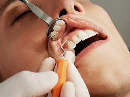
How General Dentistry Uses Imaging To Identify Early Oral Concerns
Your mouth often shows warning signs long before pain starts. General dentistry uses simple imaging to find these early oral concerns, so you can act before problems grow. A Fort Atkinson, WI dentist can use routine X-rays and photos to spot small cavities, early bone loss, and hidden infection. These tools show what the eye cannot see. They reveal changes under fillings, along the gumline, and between teeth. Early pictures help you avoid root canals, extractions, and emergency visits. They also support safer care if you have diabetes, heart disease, or a weak immune system. Regular imaging gives your dentist a clear record of your mouth over time. Then small changes stand out. You gain control. You can plan treatment that fits your life, your budget, and your health goals.
Why early imaging matters for you
Small problems in your mouth grow in quiet ways. A tiny cavity spreads through the enamel. A bit of plaque under your gum starts to break down bone. A crack in a tooth widens each time you chew.
You often feel nothing at first. You may see no stain or swelling. Yet real harm is building. That is where imaging steps in.
Dental images help your dentist:
- Find disease before it hurts
- Protect teeth and gums from major damage
- Plan treatment that is smaller, faster, and less costly
The Centers for Disease Control and Prevention explains that untreated cavities are common in children and adults. You can review current data at the CDC oral health page on tooth decay. Early imaging gives you a chance to change that story for your family.
Common imaging types in general dentistry
You see imaging as a normal part of a checkup. Each type offers a different view. Together, they give a full picture of your mouth.
Bitewing X rays
Bitewings show the crowns of your upper and lower teeth at the same time. They help your dentist see:
- Cavities between teeth
- Early bone loss from gum disease
- Changes under fillings and crowns
You usually get these every one to two years, or more often if you have a high risk for decay.
Periapical X rays
Periapical images show the whole tooth. That includes the root and the bone that holds it. Your dentist uses them to check for:
- Infection at the root tip
- Deep cracks
- Cysts or other growths near the roots
These images guide care when you have pain, swelling, or a tooth injury.
Panoramic X rays
A panoramic image wraps around your whole mouth in one picture. It shows:
- All teeth, including ones that have not come in yet
- Jaw joints and sinuses
- Wisdom teeth and their position
This type supports planning for braces, extractions, or dentures.
Digital photos
Simple photos from a small mouth camera help you see what your dentist sees. They show:
- Worn edges
- Chips and cracks
- Red or white spots on soft tissue
These photos help track change at each visit. They can also help children understand their own teeth.
How imaging finds early oral concerns
Imaging does more than confirm what your dentist already suspects. It often is the only way to see the first signs of trouble.
Tooth decay
Cavities often start between teeth. They hide under contact points where a mirror cannot reach. Bitewing X-rays show slight shadows in these spots long before a hole forms. Your dentist can treat the decay while it is still small. That helps you avoid deep fillings and possible root canals later.
Gum disease
Gum disease eats away at bone in a slow, quiet way. Early bone loss rarely hurts. Periapical and bitewing images show the height of the bone around each tooth. Your dentist can compare current images with older ones to see if the bone is dropping. Then you can start gum treatment while the teeth are still firm.
Infection and cysts
A dark ring at the tip of a root on an X-ray often means infection. A larger dark space may show a cyst. These spots may not cause pain at first. Imaging helps your dentist act before the infection spreads to other teeth or into the jaw.
Cracks and wear
Repeated stress from grinding or clenching wears teeth down. It can also lead to small cracks. Photos and X-rays together show changes in tooth shape and lines within the tooth. You can then discuss night guards or other steps to protect your teeth.
Typical imaging for different ages
Your imaging needs change as you age. The table below gives a general picture. Your dentist will adjust based on your risk and your health history.
| Age group | Common imaging | Main concerns checked |
|---|---|---|
| Children 5 to 12 | Bitewing X-rays every 1 to 2 years. Panoramic once as permanent teeth come in. | Early cavities. Tooth growth and spacing. Impacted teeth. |
| Teens 13 to 19 | Bitewing X-rays every 1 to 2 years. Panoramic for braces or wisdom teeth. | Cavities between teeth. Wisdom tooth position. Jaw growth. |
| Adults 20 to 64 | Bitewing X-rays every 1 to 2 years. Periapical as needed. Panoramic every few years. | Decay under fillings. Gum disease. Cracks and infection. |
| Older adults 65 and up | Bitewing X-rays every 1 to 2 years. Periapical for sore or loose teeth. Panoramic as needed. | Root decay. Bone loss. Fit of dentures or bridges. Oral cancer checks. |
Radiation and safety
Many people worry about X-rays. That concern is natural. Modern dental imaging uses very low radiation. Digital systems use less than older film.
Your dentist also protects you with:
- Lead aprons over your chest and lap
- Thyroid collars around your neck when needed
- Only the images that are needed for safe care
The American Dental Association explains current safety guidance at ada.org. You can review this and bring questions to your next visit.
How you can use imaging results
Images work best when you use them as a tool, not just a record. During your visit, ask your dentist to show you the pictures on the screen. Ask three simple questions.
- What looks healthy
- What concerns you right now
- What might change if I do nothing
Then talk about your options. You can choose treatment now for small issues. You can also plan steps at home, such as better brushing, more fluoride, or changes in diet.
Each new image becomes part of your health story. Over time, patterns appear. Your dentist can see if your gums stay steady, if fillings hold, and if any spots come back. You gain a sense of control and calm. You know that quiet problems are less likely to surprise you.